In This Guide
- What X-rays reveal about Hallux Rigidus staging
- When an MRI is necessary vs. an X-ray alone
- How to read your imaging report and understand what it means
- How imaging results directly shape your treatment plan
95%
diagnostic accuracy of weight-bearing X-rays for detecting hallux rigidus at Grade 2 and above, but MRI is needed to assess soft-tissue damage and cartilage loss at Grade 1
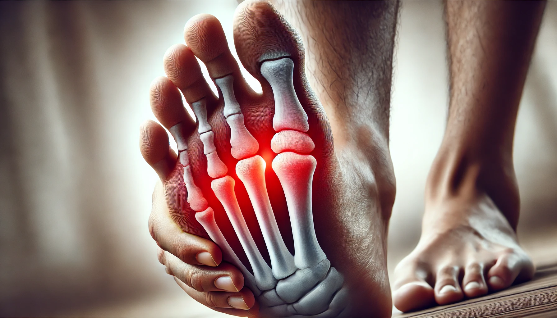
Every step you take should be effortless, but what if your big toe refused to move due to Hallux Rigidus? Understanding the roles of X-rays and MRIs is the first step toward an accurate diagnosis and an effective treatment plan.
Understanding the role of X-rays and MRIs in diagnosing this condition is essential, as early and accurate imaging helps prevent hallux rigidus. Hallux Rigidus is a progressive condition that makes even simple activities like walking painful.
Imagine waking up one day and feeling stiffness in your foot that only worsens over time. That’s the reality of Hallux Rigidus, a silent but progressive condition that can turn something as simple as walking into a painful struggle.
Untreated cases of this disease can lead to chronic discomfort and difficulty walking. Early diagnosis is key. Know more about the Early Symptoms of Hallux Rigidus.
What many people don’t realize is how much impact the right shoes can have in managing this condition. Supportive footwear is a part of your treatment. That’s why we put together a list of the 20 Best Walking Shoes for Hallux Rigidus to help you walk with less pain and greater confidence.
What Is Hallux Rigidus and Why Early Diagnosis Matters?
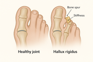
Hallux Rigidus is a form of arthritis affecting the joint at the base of the big toe, known as the metatarsophalangeal (MTP) joint. It is often caused by wear-and-tear, injury, or genetic predisposition. Its symptoms typically include pain, stiffness, and swelling when walking or standing. These symptoms vary by individual and by the degree to which their diagnosis has progressed.
Hallux Rigidus Symptoms: What to Watch For
- Painful stiffness of the big toe joint.
- Bone spurs cause an obvious bump at the top of the toe joint.
- Problems wearing shoes with narrow or rigid soles. Reduced ability to bend the toe joint upwards or downwards.
Early identification is of vital importance. Delayed diagnosis could worsen symptoms, reduce mobility, and necessitate more intensive treatments such as surgery. This is why X-rays and MRIs are crucial for diagnosing Hallux Rigidus and provide timely results.
Imaging Tests for Hallux Rigidus: X-Rays and MRIs
Doctors and healthcare providers rely heavily on imaging tools, like X-rays and MRIs, when diagnosing Hallux Rigidus. The role of X-Rays and MRIs is crucial when X-rays don’t show the full picture, helping create a precise treatment plan. The images provide a detailed view of the involved joint, giving healthcare providers greater insight into this condition.
X-rays
X-rays use electromagnetic radiation to generate images of bones and joints. They show any structural changes, such as bone spurs or narrowed joint spaces. As one of the Best Imaging Tests for Hallux Rigidus, it quickly identifies early signs, helping doctors assess severity and plan treatment. This makes X-rays often the first tool used when diagnosing suspected cases of Hallux Rigidus.
MRIs
On the other hand, Magnetic Resonance Imaging (MRI) uses strong magnetic fields and radio waves to produce detailed images of both bones and soft tissues such as cartilage and tendons. MRIs are ideal when healthcare providers require an overview of a joint.
Understanding these diagnostic methods is integral to understanding their specific roles when diagnosing Hallux Rigidus.
Understanding X-Rays’ Role in Diagnosing Hallux Rigidus
It starts with a dull ache in your big toe, nothing too serious.
You shake it off, blaming tight shoes or a long day on your feet. But soon, the stiffness sets in. Walking feels awkward, and bending your toe becomes painful. You adjust, avoiding certain movements, hoping it will fade.
But what if it doesn’t?
What if, beneath the surface, Hallux Rigidus is quietly progressing, locking your joint and making each step harder?
This is where X-ray for Hallux Rigidus Diagnosis becomes important. As one of the Best Imaging Tests for Hallux Rigidus, it provides a fast, clear picture of what’s happening inside your joint. Spotting bone spurs, joint space narrowing, and structural damage before it’s too late.
The sooner you get diagnosed, the sooner you can take control of your mobility.
X-rays are among the best and quickest methods for diagnosing Hallux Rigidus. They are fast, affordable, and highly accurate at showing any changes that have taken place to bones and joints
The Role of X-Rays and MRIs in Hallux Rigidus Diagnosis
- Identification of Joint Space Narrowing: Hallux Rigidus causes cartilage loss in the big toe joints, so x-rays show this narrowing between bones.
- Detecting Bone Spurs: Bone Spurs may indicate progression of Hallux Rigidus.
- Assess Bone Alignment: Hallux Rigidus can disrupt the alignment of bones in your foot, leading to additional discomfort.
- An X-ray for Hallux Rigidus diagnosis offers doctors an effective tool for tracking illness progression over time by comparing images. By doing this, doctors can assess whether your condition has worsened and implement any necessary treatment changes.
Limitations of X-rays:
While X-rays excel at visualizing bone structures, magnetic resonance imaging (MRI) excels at visualizing soft tissues.
Role of MRI in Diagnosing Foot Disorders
Magnetic Resonance Imaging (MRI) offers an in-depth view of soft tissues and cartilage in your foot. Employing powerful magnets and radio waves, MRI produces high-resolution images not possible with traditional X-ray techniques.
- Visualizing Cartilage Health: MRI accurately allows doctors to monitor cartilage health in joints damaged by Hallux Rigidus.
- Early Arthritis Detection: Even before symptoms become noticeable, an MRI can identify subtle signs of arthritis before any severe signs arise.
- Examining Soft Tissues: MRIs can detect inflammation in tendons or ligaments near an affected joint, helping rule out other conditions.
- Planning for Surgery: When surgery becomes necessary, an MRI provides surgeons with guidance, offering comprehensive views of affected areas.
Using both methods demonstrates why X-rays and MRIs are necessary for diagnosing Hallux Rigidus and for effective treatment. Each approach complements the other perfectly.
Limitations of MRIs:
MRIs tend to be more expensive and time-consuming than X-rays. They are reserved for cases in which additional detailed imaging is required or initial X-ray results do not provide definitive answers.
How Early Diagnosis Affects Hallux Rigidus Treatment and Mobility
At first, it’s just stiffness, a minor discomfort in your big toe.
But over time, each step feels heavier, and soon, even standing too long becomes a struggle.
What if you could stop it before it gets worse?
An early diagnosis of Hallux Rigidus is key to protecting your joints and maintaining mobility. Early diagnosis of Hallux Rigidus helps protect joints from further damage while improving overall foot health.
When identified early enough, noninvasive therapies such as custom orthotics, anti-inflammatory medication, and physical therapy are highly effective.
Patients benefit from earlier and more accurate assessments enabled by X-rays and MRIs, which help diagnose Hallux Rigidus.
Stages and Treatment Options of Hallux Rigidus:
- Stage 1 (Mild): Early symptoms, such as occasional stiffness and pain, may be managed with non-invasive therapies, including physical therapy and stretching exercises.
- Stage 2 (Moderate): Persistent issues might require injections or advanced orthotics solutions.
- Stage 3 (Severe): When joints become immovable, surgery such as joint fusion or cheilectomy may be necessary.
Early diagnosis not only lessens or avoids surgery altogether but may also reduce discomfort by maintaining mobility for as long as possible, thereby delaying or postponing the need.
The Role of X-Rays and MRIs in Regular Foot Checkups for Hallux Rigidus
You ignore the slight stiffness in your big toe, just a bad shoe day, right?
Weeks pass, then months. The discomfort turns into pain, and suddenly, even a short walk feels exhausting.
What if you had caught it earlier?
Neglecting foot pain or stiffness could result in long-term mobility challenges. So regular checkups with an orthopedic physician are incredibly important if symptoms such as pain, swelling, or limited toe movement are evident.
Diagnosis techniques for Hallux Rigidus, such as X-rays and MRIs, allow doctors to provide precise care for patients with this disease.
According to the American Orthopaedic Foot & Ankle Society and NCBI clinical resources, combining X-ray and MRI findings provides the most complete picture of hallux rigidus severity and guides appropriate treatment decisions.
Conclusion: The Role of X-Rays and MRIs in Hallux Rigidus Care
Hallux Rigidus requires careful monitoring. Ignoring symptoms like pain and stiffness in the big toe joint could result in serious consequences. Diagnostic tools such as X-rays and MRIs are the most effective imaging tests for Hallux Rigidus. They play an integral part in accurately diagnosing this condition, creating treatment plans, and ultimately improving quality of life.
Regular foot checkups are key to early diagnosis. Regular examinations help detect issues early and address them before they aggravate. Individuals experiencing persistent foot discomfort should consult healthcare providers and use advanced diagnostic tools in a proactive approach to betterment.
When combined, X-rays and MRIs can help diagnose Hallux Rigidus, enabling patients and professionals to work together toward successful results. Prioritize foot health today and begin on your journey toward healthier feet tomorrow.
Frequently Asked Questions About X-Rays and MRIs for Hallux Rigidus
Do I need an X-ray to diagnose hallux rigidus?
Yes — weight-bearing X-rays are the gold standard for diagnosing and staging hallux rigidus. They reveal joint-space narrowing, bone spur formation, and overall severity, which directly guide treatment decisions.
Imaging Note
Always request weight-bearing X-rays rather than non-weight-bearing; joint space narrowing is only visible under load and is the primary grading criterion.
What does hallux rigidus look like on an X-ray?
X-rays typically show narrowing of the first MTP joint space, osteophyte (bone spur) formation on the dorsal aspect of the joint, and, in advanced cases, subchondral sclerosis indicating significant cartilage loss.
When is an MRI needed for hallux rigidus?
An MRI is usually reserved for cases where X-rays are inconclusive, when soft tissue or cartilage involvement needs detailed assessment, or when surgery is being planned. The surgeon needs a precise map of the remaining cartilage.
Are X-rays painful or risky for diagnosing hallux rigidus?
No. Weight-bearing foot X-rays are quick, painless, and involve minimal radiation exposure. They are routinely performed in a podiatrist’s or orthopedic office with no special preparation required.
With 12 years of clinical experience specializing in degenerative joint disease, Sarah has seen the limitations of traditional medicine firsthand. As an avid marathoner, she knows that for an athlete, "rest" is a four-letter word.
At Hallux Rigidus Care, Sarah serves as the scientific anchor. She bridges the gap between sterile orthopedic literature and the practical needs of active adults.
Her mission is to provide evidence-based clarity. Sarah ensures that every piece of advice we give is medically sound, ethically grounded, and designed to keep you moving comfortably for decades to come.


